| FOOT & ANKLE |
 |
|
|
Ankle Joint:The center of the Joint is the bone called the talus.
The talus bone has a shiny joint surface covering which allows your ankle to glide effortlessly across the shiny undersurface of your large leg bone called the tibia.
When these two bones meet they form your ankle joint. On the outside of your ankle you will find a smaller, thin bone called the fibula. This bone helps prevent your major ankle bone from shifting outward.
On the other side of your ankle you will find a bump on the inside which is connected to the larger leg bone called the tibia. This part of the tibia is called the malleolus, which just means, "hammer" in Latin. |
| |
 |
| There are six major soft tissue structures that hold your ankle in place. |
|
|
-
There are six major soft tissue structures that hold your ankle in place.
-
The second most commonly torn ligament is Calcaneal-Fibular Ligament.
-
The third ligament is Posterior (meaning back) Talo-Fibular Ligament
-
The fourth ligament is the Syndesmotic Ligament, which connects the fibula to the large leg bone (tibia).
-
The soft tissue just in front of your ankle is the capsule of your ankle joint and helps keep the ankle from sliding forward along with your other ligaments.
-
The final stabilizing ligament is on the inside part of your ankle. The ligament forms a triangle shape like the Greek letter delta thus it is called the deltoid ligament. The deltoid ligament attaches the malleolus to the talus.
|
|
| |
| Foot Anatomy |
| The foot is a very complex joint with millions of combinations of movements and motions very similar to your hand. The most common form of foot pain is usually involves the heel. |
The heel bone is called the calcaneus (meaning heel in Latin). This is the largest bone of your foot and it is connected to your heel cord (Achilles tendon) above and a large broad tendon on the sole of your foot called the plantar fascia. The plantar fascia helps keep the arch of your foot supported and provides support to your foot with every step. Sometimes this ligament becomes inflamed called plantar fasciitis. This condition can cause disabling pain if it is not treated correctly. |
Sometimes heel spurs will form at the tip of the calcaneus |
| |
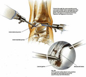
| Ankle Arthroscopy & Debridement |
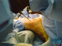
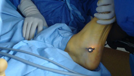
| Most ankle arthroscopy is performed under general, or occasionally regional, anaesthesia. Firstly a padded bar is placed behind the thigh. |
| Next a strap is placed around the ankle and connected to a second bar distant from the first which is attached to the operating table and allows traction to be placed across the ankle. In this way the ankle is stretched open sufficiently to allow a space with sufficient dimensions in which to operate. |
| |
 |
 |
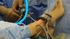
| Ankle Arthroscopy |
|
|
| |
 |
 |
| Plantar Fascia Release |
|
|
| |
 |
 |
| Most ankle arthroscopy cases can be performed as a day case surgery. |
|
| |
Return to Sport
After ankle arthroscopy you will be back to walking at 1-2 days following your ankle arthroscopy. This will be without use of a crutch. Any sporting activity is best left for at least two weeks following the procedure. Realistically, things can be built up from two weeks post-operatively but will probably take in excess of four weeks before more vigorous sporting activity will be possible. |
| |